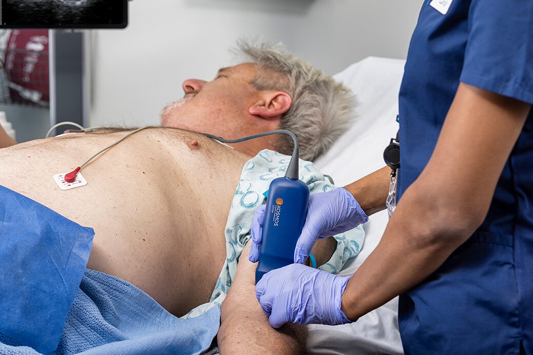
Why use ultrasound for IV access?
Placing a peripheral intravenous (PIV) catheter is a basic skill in healthcare. Millions of these procedures happen each year in various clinical settings. However, for a significant number of patients, achieving IV access is anything but routine. As many as 30% of adults and 50% of children are estimated to have difficult venous access (DVA). This can lead to multiple attempts at cannulation or the need for a central line. An estimated 35 to 50% of PIV cannulation attempts fail, which can result in complications, both immediate and delayed.
Ultrasound-guided peripheral IV (USGPIV) access offers a transformative solution. Real-time imaging allows clinicians to see target veins and guide needle placement with precision. This leads to quicker and more successful cannulations, especially in difficult cases. This article reviews the reasons for using USGPIV. It highlights key guidelines and growing evidence about the effects on patient outcomes and central line usage.
Clinical guidelines supporting ultrasound-guided IV placement
Ultrasound for vascular access has evolved from a technique reserved for specialists to the standard of care endorsed by leading healthcare organizations. Clinical guidelines increasingly recognize ultrasound as an adjunct for complex cases and a proactive solution for improving safety, efficiency, and outcomes across patient populations.
In 2021, the Infusion Nurses Society (INS) updated the 8th edition of Infusion Therapy Standards of Practice to recommend ultrasound guidance for patients with DVA, including those who have failed traditional attempts. The INS emphasizes the importance of trained personnel and image-guided insertion to improve outcomes [1].
The American College of Emergency Physicians (ACEP) encourages using point-of-care ultrasound (POCUS) for PIV access, particularly in emergency and trauma settings where rapid, reliable vascular access is critical.
The Agency for Healthcare Research and Quality (AHRQ) includes ultrasound-guided vascular access in its recommended patient safety practices, citing the role of USGIVA in reducing complications and preventing escalation to central venous access.
The American Institute of Ultrasound in Medicine (AIUM) promotes ultrasound as a core tool in vascular access protocols and supports competency-based training for clinicians.
These guidelines reflect a shift toward outcome-focused care, where real-time visualization is no longer considered optional but essential for high-quality IV access. [2,3,1]
Evidence on first-pass success and improved patient outcomes
The most compelling case for ultrasound guidance lies in the numbers. Research shows that USGPIV boosts first-attempt success rates—especially for patients with hard-to-find veins—and reduces the time, discomfort, and uncertainty of blind attempts.
Key Evidence
- A meta-analysis in the Journal of Vascular Access found ultrasound guidance improved success rates compared with traditional techniques [pooled odds ratio (OR) 3.96; 95% CI 1.75–8.94] [4].
- Another meta-analysis by Tran et al. in Ultrasound in Medicine & Biology showed a twice-higher likelihood of first-attempt success with ultrasound guidance, significantly fewer attempts at cannulation, and higher patient satisfaction [5].
- Pediatric studies echo these findings: an ED study of children with difficult access reported first-attempt success of 90% with ultrasound vs. 18% with traditional cannulation [6].
Additional Outcomes
-
Lower complication rates:
-
Frequent insertion attempts and suboptimal PIVC placement increase risks of phlebitis, thrombosis, and catheter-related infections, leading to early device failure. PIVC failure ranges from 35% to 50%, and subsequent failures are more likely after an initial failure [7]. Real-time ultrasound reduces hematomas, arterial punctures, and catheter dislodgement.
-
Improved catheter longevity:
-
Proper placement in larger, deeper veins contributes to longer-lasting access and reduced need for reinsertion [8].
-
Enhanced patient satisfaction:
-
Fewer failed attempts and less discomfort translate into higher patient-reported satisfaction and better HCAHPS scores [9].
These results support using ultrasound not only for problem-solving but also as a first-line strategy to optimize vascular access on the first try.
The role of ultrasound in reducing central line utilization
One of the most significant benefits of USGPIV is reducing unnecessary escalation from peripheral IV access to central venous catheter (CVC) placement. Central lines, while effective, carry well-documented risks, including bloodstream infections, deep vein thrombosis, and higher procedure-related costs.
The average cost of placing and managing a central line ranges from $2,000 to $5,000, not including treatment costs for complications. The CDC estimated the cost of a central line infection to be $46,000 in 2016; other studies have estimated average costs to be $17,896–$94,879. Hospitals can save hundreds of thousands of dollars annually by preventing even a small number of CVC placements [10].
Advantages of USGIVA
-
Prevention of escalation: Reliable peripheral access via ultrasound can avoid PICCs, midlines, or central lines—especially in non-critical patients.
-
Lower infection risk: Each avoided central line reduces the risk of CLABSI and improves safety metrics [11].
-
Fewer complications from repeated attempts: Given the 35–50% PIVC failure prevalence, avoiding repeated blind attempts reduces downstream failures [12,7].
-
Cost savings: An observational cohort at a tertiary center found ultrasound-trained vascular access saved approximately $83 per patient compared with traditional placement (ΔC −$83.175; p<0.001) [13].
Illustrative cases
-
A nurse-led training course for RNs on USGIVA achieved 83% first-stick success in proctored cannulations, with program costs lower than a single central line infection [14].
-
Over five years, implementing an ED ultrasound program reduced central line placement by 80%; the proportion placed in critically ill patients rose from 34% to 81% as noncritical placements fell [15].
-
In pediatrics, first-stick success reached 80% with ultrasound vs. 64% traditionally; average time decreased (6.3 vs. 14.4 minutes), and median attempts dropped (1 vs. 3) [6].
Clinical use and special populations
Ultrasound-guided IV access now spans many departments and patient groups as handheld and cart-based systems become more accessible.
Key use cases
USGIVA is widely adopted and often prioritized for patients with difficult venous access, including those with a history of IV drug use, patients on hemodialysis, or with hypotension, dehydration, edema, coagulopathy, chemoradiation, and obesity [16].
Emergency departments
Of roughly 100 million annual ED visits, nearly 60% require a PIV, and 90% of admitted patients need one. USGPIV reduces attempts and time to cannulation, helping stabilize patients faster and reduce crowding.
Oncology and hematology
Many cancer patients have fragile veins and long treatment histories. Preserving venous access with well-placed peripheral lines is critical.
Obese and bariatric patients
With increased adipose tissue, veins are harder to find. Ultrasound reveals viable vessels hidden beneath the surface.
Pediatrics and neonatology
Children have smaller, less visible veins and may experience greater anxiety with multiple attempts. Real-time imaging makes IV access faster, safer, and less traumatic.
Pre-op and anesthesia
Reliable IV access is a prerequisite for surgery and anesthesia. Ultrasound ensures timely access without resorting to more difficult procedures.
Looking ahead: next steps
Ultrasound-guided IV access is more than a procedural upgrade—it’s a paradigm shift in patient care. The evidence is clear: this approach increases success, decreases complications, and prevents the cascade of interventions that too often follow multiple failed attempts at venous access.
Multiple professional societies endorse USGIVA, and the evidence reveals significant benefits. Moreover, clinicians who adopt ultrasound for PIV rarely look back. Whether managing a high-acuity emergency department or supporting patients through long-term treatment, USGPIV can elevate the quality and consistency of care.
Let’s talk about implementation
Curious about how to get started—or how to expand an existing ultrasound-guided IV program?
Our team can help with:
- Device selection and comparison
- Training options
- Workflow optimization
- Scaling across multiple departments
Ask us today for more information on implementing a cost-effective USGIVA program at your facility.